
- St. Olaf News
- Features
- For the Media
- Media Relations
- Kari VanDerVeen
- Alumni Hall
- 507-786-3970 office
- 507-412-1036 cell
- vanderve@stolaf.edu
 RSS Feed
Note: This article is over a year old and information contained in it may no longer be accurate. Please use the contact information in the lower-left corner to verify any information in this article.
Of Tetrahymena thermophila and tomograms
February 24, 2012
Like many visitors to Colorado, a group of 10 St. Olaf students recently enjoyed visiting museums, skiing, snow-boarding, and snow-shoeing in the Rockies. But unlike most visitors to the "Centennial State," they were spending most of their time using electron microscopy in an elite national facility: the Boulder Laboratory for 3-D Electron Microscopy of the Cell.
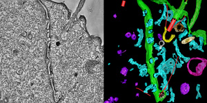 |
| A cell as seen through an electron microscope (left) and the same view after modeling by students. See more extensive modeling in the video below. Images courtesy of Trevor Romsdahl '12. |
The students were enrolled in the Interim Electron Microscopy course led by Professor of Biology Eric Cole. During the month of January, the group explored a gallery of modern microscopy techniques that culminated in a week-long training session at the facility — one of only four in the country that supports public use of the technology utilized by the students.
"What makes our experience unique is that the facility took in a group composed entirely of undergraduates from a liberal arts college, and devoted a week to train us and grant access to their instruments," explains Cole. Typically, applicants for the use of the facilities are researchers from universities — not small liberal arts colleges like St. Olaf — that have received grants from the National Institutes of Health to research cell biology. But the lab liked the idea Cole proposed of bringing a class to Boulder after they taught him 3-D tomography in 2008.
"We worked with machines that most graduate-level researchers don't even get to use," says Tom Mork '13. "We took full advantage of the opportunity."
Learning the ropes
The facility spent many hours preparing for the students, who in turn conducted extensive research to prepare themselves. During the first week of the course, students learned the biology of a single-celled organism, Tetrahymena thermophila. During the second week, students traveled to the University of Colorado Boulder, where they first learned conventional electron microscopy before using technological principles applied to the electron microscope to generate a 3-D computer tomogram (basically a CT scan) of a volume of cellular material. After each pair of students generated a tomogram, the class spent the next two weeks modeling the various cellular components and creating presentations that included animations of the materials.
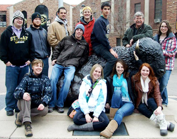 |
| The class included (front row, l–r): St. Olaf Research Associate Erica Zweifel, who helped St. Olaf Professor of Biology Eric Cole lead the students; Tom Mork '13; Kristine Elwood '12; Natasha Seliski '12; and Gretchen Becer '13. Back row: David Loe '14, Trevor Romsdahl '12, Matt Lefebvre '12, Tyler Aronstein '12, Steve Kannenberg '13, Cole, and Anna Ballard '13. |
The experience certainly made an impact on the students.
"As opposed to the traditional microscope images we're used to seeing, the 3-D models we built from our data allowed us to observe and analyze this cellular phenomenon in a much more detailed and holistic way," says Anna Ballard '13.
"They look so cool — words do not do [the images] justice," adds Natasha Seliski '12. "But the highlight of the trip was being with such a dynamic group of individuals who all shared the same passion for scientific knowledge and understanding."
Age of discovery
Not only were the students subjected to a one-of-a-kind experience, they also made several discoveries while they were at it, including the chance to confirm earlier studies showing that tiny pores form in the membranes that separate the two mating cells. The pores somehow expand to allow the nuclei to pass through. "Second, we discovered that these individual 'pores' are each filled with a delicate branching membrane system never before seen," says Cole.
While the trip was originally planned as a one-time class, the lab director has invited Cole to return with a group in the future. And Eileen O'Toole, a research associate at the laboratory, was impressed enough with the students she had worked with to send Cole the following note:
"This was a wonderful day and I’m stunned with the dedication and focus your students have. I have never had a group like this in the 20+ years I’ve been teaching. Congrats to you for having such a special group of students."
The students even impressed their teacher. "When they presented their final work it took my breath away," Cole says. "They had put in dozens of hours and accomplished so much. I was humbled and moved beyond words."
Read what Scientific American had to say about this research.
In the video above, students began by taking thin slices from a single cell (300 nm thick). These slices were then photographed through an electron microscope from 240 different angles. A computer then built a 3-D reconstruction of the intracellular-space called a tomogram (a 3-D electron microscope image similar to the imaging created by CT-scan). After building the tomogram, a color-model was rendered with the electron tomogram in the background. Such 3-D models can then be explored from any angle or vantage point. This video wanders though a landscape of microscopic membranes and cytoskeletal fibers that are associated with the nuclear exchange junction that forms between mating freshwater protozoan Tetrahymena cells.